

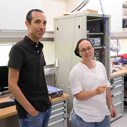
In a stunning leap forward for imaging technology, researchers at the Weizmann Institute of Science have developed a revolutionary form of nano-MRI capable of visualizing structures at the scale of a single nanometer—one-billionth of a meter. This innovation, led by Dr. Amit Finkler and his team in the Chemical and Biological Physics Department, could transform how scientists study molecules, materials, and even future pharmaceuticals.
Traditional MRI, widely used in hospitals, can resolve details down to a tenth of a millimeter. But for researchers aiming to study the atomic structure of individual molecules, that’s not nearly enough. The Weizmann team’s new device uses a synthetic diamond embedded with a nitrogen-vacancy center—a tiny defect that acts as a highly sensitive magnetic sensor. This allows the system to detect the faintest magnetic signals from particles as small as 50 nanometers away.
What sets this nano-MRI apart is its ability to operate at room temperature and under real-world conditions, making it a practical tool for studying materials as they function. By combining a confocal microscope with an atomic force microscope, the researchers achieved unprecedented resolution, potentially enabling scientists to map out the atomic structure of molecules in exquisite detail.
This breakthrough not only cements the Weizmann Institute’s reputation as a global leader in cutting-edge research but also opens new doors in materials science, drug development, and nanotechnology. As Dr. Finkler’s team continues to refine the technology, the dream of seeing the invisible world of molecules in real time is becoming a reality.
A version of this article was originally published at Weizmann Wonder Wander, and picked up at Phys.org.